
JSER Policies
JSER Online
JSER Data
Frequency: quarterly
ISSN: 1409-6099 (Print)
ISSN: 1857-663X (Online)
Authors Info


- Read: 71203
|
УШЕРОВИОТ СИНДРОМ ВО ОБРАЗОВНAТA СРЕДИНА: ИЗВОРНИ СТРАТЕГИИ ЗА ИДЕНТИФИКАЦИЈА РАЗВИЕНИ ВО ИНДИЈА
Гнанатикам Викторија НАОМИ
Институт за домашна наука и високо образование за жени „Авинашилингам“ |
|
USHER SYNDROME IN EDUCATIONAL SETTINGS: INDIGENOUS IDENTIFICATION STRATEGIES DEVELOPED IN INDIA
Gnanathicam Victoria NAOMI
Avinashilingam Institute for Home Science and Higher Education for Women – |
|
Примено: 13. 05. 2013 |
|
Recived: 13. 05. 2013 |
|
|
|
|
|
|
|
|
|
Вовед |
|
Introduction |
|
|
|
|
|
Лицата кои се вродено глуви или наглуви може да се соочат со постепено губење на нивниот вид во доцното детство, а комбинацијата на овие две загуби резултира во една од најпредизвикувачките нарушувања за една индивидуа: состојба наречена Ушеров синдром, кој пак е најчест предизвикувач на глуво-слепост кај возрасни (1–3). Ушеровиот синдром е генетско нарушување кое вклучува губење на слухот и ретинална пигментоза (РП), прогресивна дегенеративна болест на окото (4–6). Губитокот на способноста за слушање и ретиналната пигментоза се двата главни критериуми со кои се карактеризира Ушеровиот синдром. Ретиналната пигментоза е вродено нарушување што доведува до постепено дегенерирање на фоторецепторите (стапчињата) во мрежницата (5 - 7, 8). Периферната мрежница е нападната прва. Во периферната мрежница се концентрирани најголем број стапчиња - фоторецепторни клетки (150 милиони) кои му овозможуваат на лицето да гледа при замрачување. Стапчињата се одговорни за ноќно гледање и детекција на надворешно движење (9–18). Кога стапчињата се нападнати, лицето може да има неизедначен и нерамномерен поглед (скотома: црна дамка), круг во кој недостасуваат информации (кружна скотома). Лицето може да има тунелен вид (надворешните, долните и горните полиња се намалени). Лицето се чувствува како да гледа низ пиштол. На тој начин ретиналната пигментоза предизвикува ноќно слепило, обично со загуба на периферниот вид (19–24). Превеленцијата на РП е околу 1:4000 со еднонасочен подвид како најчеста појава (25–30). Иако само 4 во 100 000 лица се дијагностицирани со Ушеров синдром, се претпоставува дека 3 до 6 проценти од луѓето кои имаат наследно губење на слухот го имаат овој синдром. Во развиените земји како САД, 4 бебиња од 100 000 новороденчиња имаат Ушеров синдром. (31, 32). |
|
People who are congenitally deaf or hard of hearing may experience gradual loss of their sight in late childhood, and the combination of these two losses results in one of the most challenging disabilities for individuals: a condition called Usher Syndrome, which is the most common cause for deaf-blindness in adults (1–3). Usher Syndrome is a genetic disorder involving both hearing loss, and retinitis pigmentosa (RP), a progressive degenerative eye disease (4–6). The loss of the ability to hear and retinitis pigmentosa are the two major criteria that characterize Usher Syndrome. Retinitis pigmentosa is an inherited disorder that results in gradual deterioration of the light receptor cells (rods) in the retina (5–7, 8). The peripheral retina is affected first. The peripheral retina contains the greatest concentration of rod cells (150 million), which allows a person to see in dim light. The rods are responsible for night vision, and they detect outer movement (9–18). Once the rods are affected, a person may have spotty or patchy vision (scotoma: blind spot), a ring of missing information (ring scotoma). The person may have a tunnel vision effect (outer, lower, and upper fields are decreased). The affected person feels as if he is seeing through a gun. Thus, retinitis pigmentosa causes night blindness usually with the loss of peripheral vision (19–24). The prevalence of RP overall is about 1:4000 with the simplex subtype being the most common (25–30). While only approximately 4 in 100,000 people are diagnosed with Usher Syndrome, it is estimated that 3 to 6 percent of people who have a hereditary hearing loss have the syndrome. In developed countries such as the United States, about 4 babies in every 100,000 births have Usher Syndrome (31, 32). |
|
|
|
|
|
Знаци на Ушеров синдром |
|
Signs of Usher syndrome |
|
|
|
|
|
Иако многу ученици со Ушеров синдром не се свесни дека имаат губиток на видот, тие сфаќаат дека имаат потешкотии во гледањето што останатите лица ги немаат. Првиот и најчест знак на Ушеровиот синдром е неможноста за гледање ноќе. Вториот знак е неможноста да се гледа периферно (горе, долу и настрана) во услови на осветленост. Ученик со Ушеров синдром може да не забележи дека друг ученик го поздравува отстрана. Друг знак е потешкотија во прилагодувањето на променливо осветлување: влегување или излегување од зграда на сонце или влегување/излегување во/од затемнета просторија. Дополнителни знаци се проблемите со одржување на рамнотежата и чувствителност на блесок (4–6). Затоа, тестирањето е само првиот чекор при идентификацијата на Ушеровиот синдром. Тестирањето е важно затоа што раната идентификација влијае на медицинската одлука, на одлуките во врска со образованието и услугите, како и социјалните импликации за ученикот и неговото семејство (33). Во голем број училишта во земјите во развој не се прави тестирање за ретинална пигментоза, што може да доведе до неидентификување на Ушеровиот синдром. Повеќето од наставниците на ученици со слушно и визуелно оштетување треба уште да се запознаваат со преваленцијата и карактеристиките на Ушеровиот синдром. Лекарите немаат доволно знаење за ова нарушување. Речиси и не постои клиничка дијагностика која е достапна во државата за идентификација на Ушеровиот синдром. Земајќи ги предвид овие фактори студијата беше испланирана со следните цели:
|
|
Although many students with Usher Syndrome may be unaware that they have a visual loss, they do realize that they have difficulties with vision that other persons do not seem to have. A common first sign of Usher Syndrome is the inability to see clearly at night. The second sign is the inability to see peripherally (above, below, and to the side) under any lighting conditions. A student with Usher Syndrome may not notice another student waving hello from the side, and obstacle on the floor etc. Another sign is difficulty in adjusting to changes in lighting: entering or leaving a building on a bright sunny day or entering / leaving a darkened room. Additional signs are problems in maintaining balance and sensitivity to glare (4–6). Therefore, screening is only the first step in identifying Usher Syndrome. Screening is important because early identification impacts medical decisions, education decisions / services, and social implications for the student as well as the family members (33). Vision screening for retinitis pigments is not performed in a school setting in many developing countries, which may result in an under-identification of students with Usher Syndrome. Most of the teachers of the hearing and the visually impaired children in India don’t have enough knowledge about the prevalence and implications of Usher Syndrome. The medical professionals have little knowledge of this disorder. There is hardly any clinical diagnosis available in the country for identification of Usher Syndrome. Keeping these factors in mind the study was planned with the following objectives:
|
|
|
|
|
|
Метод |
|
Method |
|
|
|
|
|
Област |
|
Area |
|
Студијата беше спроведена во локално училиште за глуви, локално училиште за слепи и инклузивно училиште во областите Којмбаторе и Салем во Тамил Наду. Во студијата беа вклучени пет локални училишта за глуви, две локални училишта за слепи и 21 образовна програма за ученици со попреченост. |
|
The study was conducted in the residential school for the deaf, the residential school for the blind and the Inclusive schools in Coimbatore and Salem districts of Tamil Nadu. The study sample included 5 Residential schools for the deaf, 2 residential schools for the blind and 21 inclusive education programs for the disabled. |
|
|
|
|
|
Примерок |
|
Sample |
|
Вкупно 9793 ученици беа тестирани, од кои 695 беа од локалните училишта и 98 беа дел од инклузивните образовни програми. Пред да се спроведе тестирањето, беше спроведено интервју со класните раководители, старателите и врсниците (оние кои се блиски до учениците) за да се открие дали некој од учениците има проблеми со видот, по изглед, по однесување или по жалбите за користење на листи за проверка за тестирање на видот. Овој вид на групно тестирање беше спроведен за да се елиминираат оние со нормален вид. Во инклузивното училиште беа идентификувани 12 ученици со РП од секундарните медицински податоци достапни во училиштето, а ниту еден од нив немаше проблеми со слухот. Писмена дозвола да се имплементира студијата во инклузивниот образовен систем беше добиена од директорот за образование на Којмбаторе и Салем и од локалниот директор на социјалната служба за лица со попреченост на Којмбаторе и Салем за локалните училишта за глуви и слепи. |
|
A total of 793 students were screened, out of which 695 students were from residential settings, and 98 from the inclusive education program. Prior to formal screening, an interview was conducted involving the classroom teachers, caretakers and peers (those who are closer to the students) to find out whether any student had any visual problem by appearance or behavior or complaints using the vision screening checklist. This type of mass screening was conducted to eliminate those with normal vision. In the Inclusive Education for the disabled, the investigators identified 12 RP students from the secondary medical data available in the school and none of them had hearing defect. Written permission for implementation of the study was obtained from the Chief Education Officer of Coimbatore and Salem districts, and from District Disabled Welfare Officer of Coimbatore and Salem districts. |
|
|
|
|
|
Алатки за тестирање: |
|
Screening tools: |
|
Лицата со Ушеров синдром беа изложени на намелена визулена бистрина, ограничување на видното поле (34,35), недоволна адаптација на темно, светло (36–39), блесок (40), чувствителност на контраст (41–42) и проблеми во рамнотежата (43). Со цел да се идентификуваат овие тешкотии кај децата со оштетен слух во оваа студија беа развиени изворни направи и техники, како и едноставни направи за тестирање и проценка на далековидоста и кратковидоста, како и на видното поле кои веќе беа тестирани. Тестирањето беше направено за да се идентификуваат оние ученици кај кои постои ризик од Ушеров синдром. |
|
Persons with Usher Syndrome experience reduced visual acuity, field restriction (34,35), lack of dark light adaptation (36–39), glare (40) and contrast sensitivity (41–42) and balance problems (43). In order to identify these major difficulties in hearing impaired children, indigenous devices and techniques have been developed in the study, as well as simple screening devices for assessment of distance and near vision and field of vision which were already tested. The screening was done to identify those individuals who were at risk for Usher Syndrome. |
|
|
|
|
|
I. Далековидост и кратковидост |
|
I. Distance vision and near vision |
|
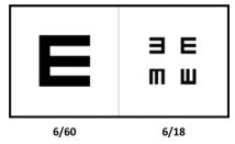
Е-табелата беше користена за да се оцени далековидоста. Само одредени нивоа беа тестирани, затоа што ова беше положи/падна тест за секое ниво. Keffee (44) го разви приборот за мерење на далековидоста и видното поле за земјите во развој, и овие тестови беа користени во студијата. |
|
A Tumbling E card was used to assess distance vision. Only specific levels were tested, as this was a pass/fail test for each level. Keffee (44) developed the assessment kit for measuring distance vision and field of vision for developing countries, and these tests were used in the study. |
|
Слика 1:Кратковидост и далековидост |
|
Figure 1: Distance vision and near vision |
|
|
|
|
|
Ако бистрината е 6/18 или подобра, видот се смета дека е во нормални рамки. Ако е 6/60 или 3/60, се смета дека лицето има намален вид. Ако најголемиот симбол не може да се види на оддалеченост од 3 метри, се смета дека бистрината е помала од 3/60 и, според дефиницијата на СЗО (1992), лицето се смета за слепо. |
|
If the acuity is 6/18 or better, the vision is considered within normal range. If acuity is 6/60 or 3/60, the person is categorized as having low vision. If the largest symbol cannot be recognized at 3 meters, acuity is recorded as less than 3/60 and the participant is regarded as blind according to the WHO (1992) definition (59). This test card was used to measure distance vision. |
|
|
|
|
|
Табела за кратковидост |
|
Near vision card |
|
II. Видно поле |
|
II. Visual field |
|
|
|
|
|
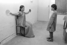
Тестот за конфронтација беше користен за да се оцени ограниченото или намалено видно поле на индивидуата. Губењето на видното поле со ретиналната пигментоза е прогресивна состојба што се развива бавно. Лицето што се тестира стои на 0,5 метри од оценувачот. Овој едноставен тест за видно поле може да се направи така што ученикот ќе гледа напред, во очите на оценувачот, и од него ќе се бара да ја подигне раката кога ќе ги види прстите или некој светол предмет како диск во раката на оценувачот што се движи отстрана. |
|
The confrontation test was used to assess the restricted or reduced peripheral visual field of an individual. The field loss associated with retinitis pigmentosa is a progressive condition which develops slowly. The person being tested stands at 0.5 meters away from the evaluator. This simple visual field test can be done by having the student look straight ahead, watching the evaluator’s eyes and asking the person to raise his/her hand when he/she sees the fingers or some bright objects like a disc in the evaluator’s hand moving from the side. |
|
Слика 2:Тест за конфронтација |
|
Figure 2: Confrontation test |
|
|
|
|
|
Ако субјектот не успее да ја види раката на другото лице како му мавта отстрана, тоа лице се смета дека има губиток на видното поле. Нивото на губиток на видното поле се смета за мало, средно или значително во десната, левата, горната или долната позиција од очите. Ако видното поле е неоштетено во периферијата, се смета дека студентите се ризични затоа што импликацијата на РП е губење на периферниот вид. Ако периферниот вид е сериозно намален, се смета за значително губење на видот, а ако е минимално, тогаш се смета за мал губиток на видното поле. Во тестот, 10 ученици беа идентификувани дека имаат периферен губиток на видот и понатаму беа испитувани за адаптација во мрак, блесок и чувствителност на контраст. |
|
If the subject fails to glance at another person's hand waving from the side, that individual is considered to have a visual field loss. The amount of the visual field loss is described as being slight, moderate or significant in right or left, upper or lower positions from the eyes. If the visual field is not intact at the periphery, students are considered at risk because the implication of RP is peripheral vision loss. If the peripheral vision is severely restricted, it is described as a significant vision loss, and if it is minimally restricted, it is considered as slight. In the test, 10 students were identified as having a peripheral vision loss and they were further assessed for their dark adaptation, glare and contrast sensitivity. |
|
|
|
|
|
III. Адаптација во мрак |
|
III. Dark adaptation |
|
|
|
|
|
Ретиналната пигментоза обично започнува да се манифестира како неможност за гледање при замрачување. Тогаш е неопходно тестирањето за адаптација во мрак. |
|
Retinitis pigmentosa usually begins to manifest itself as an inability to see well in dim lighting. Therefore, a dark adaptation screening is crucial. |
|
|
|
|
|
IV. Чувствителност на блесок |
|
IV. Glare sensitivity |
|
|
|
|
|
Тестот се користи за да се открие дали лицето има проблем во прилагодувањето кога од дневна светлина ќе влезе во затемнета просторија. Ако кај лицето се забележи тромавост кога ќе се соочи со светлина, се смета дека тоа е индикација за чувствителност на блесок. Овој тест покажува дали таквите лица имаат тешкотии во приспособувањето на нивниот вид. |
|
The test was conducted to discover whether the participants had a problem in adjusting their vision when coming from bright sunlight to a darkened place. If participants exhibited clumsiness while facing a bright light, this may be interpreted as an indication of glare sensitivity. This test indicates whether they have difficulty adjusting their vision. |
|
|
|
|
|
V. Чувствителност на контраст |
|
V. Contrast sensitivity |
|
|
|
|
|
Чувствителноста на контраст беше оценувана со два теста: |
|
The contrast sensitivity was assessed by two tests |
|
|
|
|
|
1. Букви со мал контраст и повторување на линија со мал контраст – од учениците се бара да ги поврзат точките или да ја повторат линијата со мал контраст за да го добијат обликот и да ја прочитаат картичката со букви со мал контраст. |
|
1. Low contrast letters and ditto copying - the students were asked to join/copy a low contrast dotted line to make the shape or picture and read the low contrast letter card. |
|
Слика 3 Повторување на линија |
|
Слика 4Букви со мал контраст |
|
Figure 3 Ditta coping |
|
Figure 4 Low contrast letters |
|
|
|
|
|
Ако не успеат да ги поврзат точките или да ја повторат линијата со мал контраст или ако им е потребен поголем контраст за да успеат, ова би бил индикатор за можна РП за која се потребни поголем контраст и подобра осветленост. |
|
If they failed to join or copy the low contrast line and needed a higher contrast line in order to be successful, this would be an indicator of the possibility for RP for which high contrast and good illumination are required. |
|
Слика5 Пресипување вода во осветлена соба |
|
Слика 6Пресипување вода во затемнета просторија |
|
Figure 5 Pouring water in illuminated room |
|
Figure 6 Pouring water in dim room |
|
|
|
|
|
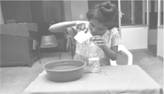
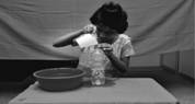
Учесниците беа седнати во слабо осветлена соба за да ги изведат овие задачи. Шишето со вода е безбојно исто како и водата. Нема разлика помеѓу овие две и затоа се сметаат за предмети со мал контраст. Неможноста да се изврши задачата со мал контраст се смета за индикатор за постоење на ретинална пигментоза. Активноста беше извршена во добро осветлени и во замрачени простории и беше споредено извршувањето на активноста во однос на разлевањето и квалитетот на извршување, на пример држењето на чашата и визуелното однесување како на пример, колку близу се очите до предметот. |
|
The participants were seated in a dimly illuminated room to perform these tasks. The water bottle is colorless like the water. There is no distinction between these two and hence, they are considered as low contrast items. Being unable to perform the task with less contrast would be considered to be indicative of the possibility for retinitis pigmentosa. The activity was done in high illumination and dim lighting and the performances were compared in terms of spilling and quality of performing, e.g. holding of the cup, and visual behavior such as how close the eyes were to the object. |
|
|
|
|
|
VI. Чувство на рамнотежа |
|
VI. Sense of balance |
|
|
|
|
|
Проблемите со рамнотежата беа тестирани на тој начин што субјектите беа замолени да седнат во темна просторија. Познато место беше модифицирано со поставување на мебел, предмети и лица околу него/неа за да се открие дали тој/таа покажува вознемиреност при движењето во нова средина. Испитувачите ги набљудуваа учениците во светли и темни ситуации и потоа набљудуваа дали во темна просторија тој/таа ја губи својата рамнотежа. Губењето на рамнотежата беше критериум за паѓање на тестот. |
|
Balance problems were tested by asking the subject to sit in a dark place. The familiar place was modified with the placement of some furniture, things and people around him/her to find out whether s/he showed anxiety to move in the new environment. The investigators observed the student in bright and dark situations, and then observed whether he/she lost his/her balance in a dark place. Losing balance was considered as a criterion for failing. |
|
|
|
|
|
Функционална визуелна проценка |
|
Functional vision assessment |
|
|
|
|
|
Функционално визуелните способности на учениците беа оценувани за да се одреди колку тие можат да го употребуваат својот вид за секојдневни активности. Проценката не тестираше РП, туку проценуваше колку индивидуата го користи својот вид за визуелни задачи. Следниве области беа оценувани со помош на процедурата за функционалноста на видот на Keffee (44). |
|
Functional visual skills of the students were assessed to determine how much they could use their vision for day to day activities. The assessment did not specifically screen for RP; it assessed how much individuals used their vision for visual tasks. The following areas were assessed using Keffee’s (44) functional vision assessment procedure. |
|
|
|
|
|
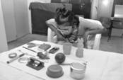
a. Визуелно следење |
|
а. Visual tracking |

|
Слика 7Следење на топка |
|
Figure 7 Tracking ball |
|
|
|
|
|
б. Визуелно скенирање |
|
b. Visual scanning |
|
Слика 8Скенирање предмети |
|
Figure 8 Scanning objects |
|
|
|
|
|
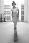
в. Визуелна моторика |
|
c. Visual motor |
|
Слика 9 Одење по тули |
|
Figure 9Walking on bricks |
|
|
|
|
|
Се набљудуваше дали лицето покажува визуелно моторна координација за да одржи рамнотежа. Додека се движи во средината, се користи периферниот вид за скенирање и следење за да види што има во средината и да овозможи координиран од. Ако субјектот не може да се движи координирано во средината, се смета дека тој/таа паднале при оваа процена и дека кај нив постои ризик за постоење РП. |
|
Observations were made whether the person demonstrated visual motor coordination to maintain balance. While moving in an environment, peripheral vision is used for scanning and tracking what is in the environment, enabling a coordinated gait. If the subject was unable to move in a coordinated fashion in this environment, he/she wеre considered to have failed this assessment, and to be at risk of having RP. |
|
Резултати |
|
Results |
|
|
|
|
|
Резултат 1: |
|
Result 1: |
|
|
|
|
|
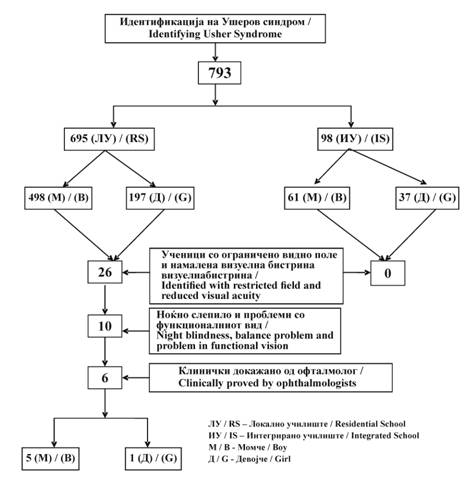
Сликата 10 го опишува тестирањето за ризик за постоење Ушеров синдром. Ова ја покажува елиминацијата на учесниците фаза по фаза сè до последниот учесник кај кој постои ризик за Ушеров синдром. |
|
Figure 10 shows describes the screening for students at risk of having Usher Syndrome. This shows the stage by stage elimination of participants and at the last subjects to risk of having Usher Syndrome. |
|
Слика 10: Идентификација на Ушеров синдром |
|
Figure 10:Identification of Usher Syndrome |
||
|
|
|
|
||
|
Резултат 2: Бракови со вкрстено сродство меѓу родителите |
|
Result 2: Consanguineous marriage between parents |
||
|
Ушеровиот синдром е наследен, поминува од родителите на нивните деца преку гените. Резултатите од студијата покажуваат дека 90% од учениците кај кои постоеше ризик за Ушеров синдром имаат родители кои се во некакво сродство. Сите родители имаа нормален слух и вид, но не знаеја дали се носители на мутираните гени на Ушеровиот синдром. |
|
Usher Syndrome is inherited, passing from parents to their children through genes. The present study presents evidence that 90% of the students who were at risk of having Usher Syndrome had parents who were closely related. All of these parents had normal hearing and vision but, did not know if they were carriers of an Usher Syndrome gene mutation. |
||
|
|
|
|
||
|
Резултат 3: Видно поле |
|
Result 3: Visual field |
||
|
Лицата со Ушеров синдром имаат проблеми со видот. Кај овие деца проблемите со видот се јавуваат обично околу десеттата година. Бројни студии што го користеле овој метод откриле дека половина век од контракцијата на видното поле (времето кога половина од видното поле е изгубена) останува константен кај сите видови РП околу пет до осум години (44–45). Проблемите со видот обично започнуваат со тешкотии со гледањето ноќе и се развиваат брзо сè додека лицето не остане сосема слепо. Анализите за ограничувањето на видното поле кај 10 посебни случаи откриле дека кај 80% од нив постои ограничување на видното поле. |
|
The persons with Usher Syndrome have visual problems. These children usually begin to develop vision problems by the age of ten. A number of studies using this method have determined that the half-life of visual field contraction (the time over which half of the remaining field area is lost) appears to remain remarkably constant across all types of RP at about five to eight years (44–45). Visual problems most often begin with difficulty seeing at night and tend to progress rapidly until the individual becomes completely blind. An analysis of the field restriction of 10 special cases found that 80% of them had field restriction. |
||
|
|
|
|
||
|
Резултат 4: Визуелна бистрина |
|
Result 4: Visual acuity |
||
|
|
|
|
||
|
Резултат 5: Адаптација на мрак |
|
Result 5: Dark adaptation |
||
|
|
|
|
||
|
Резултат 6: Чувствителност на блесок |
|
Result 6: Glare sensitivity |
||
|
|
|
|
||
|
Резултат 7: Чувствителност на контраст |
|
Result 7: Contrast sensitivity |
||
|
|
|
|
||
|
Резултат 8: |
|
Result 8: |
||
|
|
|
|
||
|
Дискусија |
|
Discussion |
||
|
Оваа студија покажа дека децата со Ушеров синдром може да бидат дијагностицирани во училишната средина. Авторите идентификуваа шест случаи со Ушеров синдром во училишната средина со користење на едноставни техники. |
|
This study demonstrated that children with Usher Syndrome may be unidentified in educational settings. The authors identified six cases with Usher Syndrome in the educational setting using simple techniques. |
||
|
|
|
|
||
|
Конфликт на интереси |
|
Conflict of interests |
||
|
Авторите изјавуваат дека немаат конфликт на интереси. |
|
Author declare no conflict of interest. |
||
|
|
|
|
||
|
Благодарност |
|
Acknowledgement |
||
|
Ние би сакале да ја изразиме нашата благодарност до генералниот директор за образование на областите Којмбаторе и Салем на Тамил Наду за нивната дозвола да се спроведе истражувањето за инклузивните образовни програми и на окружниот директор за благосостојбата на лицата со попреченост на Којмбаторе и на областа Салем за дозволата за училиштата за глуви и училиштата за слепи. Ние би сакале да им се заблагодариме на властите, раководителите, наставниците и воспитувачите во посебните училишта за олеснување на спроведувањето на оваа студија. Длабоко им се заблагодаруваме на Srinivasan G. Rao, консултант-офталмолог, Очната болница Vasan, Coimbatore за медицинската дијагноза на ретинитис пигментоза. И, на крај, голема благодарност на членовите на различните семејства за нивното трпение и соработка во ова истражување. |
|
We would like to express our gratitude to the Chief Educational Officer of Coimbatore and Salem districts of Tamil Nadu for their permission to conduct the study in the inclusive education programmes and the District Disabled Welfare Officer of Coimbatore and Salem districts for permission for the Residential School for the Deaf and the Residential School for the Blind. We would like to thank the authorities, heads, teachers and caretakers of special schools for facilitating the conduct of this study. We gratefully acknowledge Dr. Srinivasan G. Rao, Consultant Ophthalmologist, Vasan Eye Care Hospital, Coimbatore for the medical diagnosis for retinitis pigmentosa. Finally many thanks to the members of the various families for their patience and persistent cooperation in this research. |
||
|
|
|
|
||
|
Citation: Naomi VG. Hemambigai C. Usher Syndrome in Educational Settings: Indigenous Identification Strategies Developed in India. J Spec Educ Rehab 2013; 14(3-4):115-132. doi: 10.2478/JSER-2013-001410.2478 |
||||
|
Article Level Metrics |
||||
|
Литература / Reference: |
|
|
||
|
|
|
||
|
|
|
|
||
Follow Us
Share Us
Journal metrics
-
 SNIP 0.059
 -
IPP 0.07
-
 SJR 0.13
 -
 h5-index 7
 -
 Google-based impact factor: 0.68
Indexed in


![]()
10 Most Read Articles
- PARENTAL ACCEPTANCE / REJECTION AND EMOTIONAL INTELLIGENCE AMONG ADOLESCENTS WITH AND WITHOUT DELINQUENT BEHAVIOR
- RELATIONSHIP BETWEEN LIFE BUILDING SKILLS AND SOCIAL ADJUSTMENT OF STUDENTS WITH HEARING IMPAIRMENT: IMPLICATIONS FOR COUNSELING
- EXPERIENCES FROM THE EDUCATIONAL SYSTEM – NARRATIVES OF PARENTS WITH CHILDREN WITH DISABILITIES IN CROATIA
- INOVATIONS IN THERAPY OF AUTISM
- DIAGNOSTIC AND TREATMENT OPTIONS IN AUTISTIC SPECTRUM DISORDERS – AN OVERVIEW
- AUTISM AND TUBEROUS SCLEROSIS
- REVIEW OF SPECIAL EDUCATION PROGRAMS IN JORDAN: CURRENT PRACTICES, CHALLENGES, AND PROSPECTS
- THE DURATION AND PHASES OF QUALITATIVE RESEARCH
- REHABILITATION OF PERSONS WITH CEREBRAL PALSY
- DISORDERED ATTENTION AS NEUROPSYCHOLOGICAL COGNITIVE DISFUNCTION